
Skin cancer is a widespread and potentially life-threatening condition. McCollough Plastic Surgery offers various methods to remove and destroy skin cancer cells, including the cutting-edge MOHS Micrographic Surgery, widely hailed as the most accurate method of removing skin cancers and preserving the most adjacent tissue. Trust our expert care for skin cancer in Gulf Shores, AL.
The Risk of Skin Cancer
Repeated and prolonged exposure to ultraviolet light can be damaging to our skin. This can come from either sun exposure or tanning beds. The effects of these damaging rays are cumulative over our lifetimes. The more exposure we get, the higher our chances of eventually getting some form of skin cancer. The fairer the complexion, the greater the risk, whereas dark-skinned or black individuals have a much lower risk. Suntan lotions with a sun protection factor greater than 15 can help protect the skin and should always be used prior to any sun exposure and reapplied frequently.
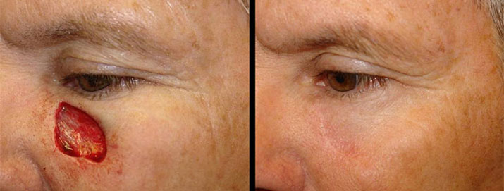
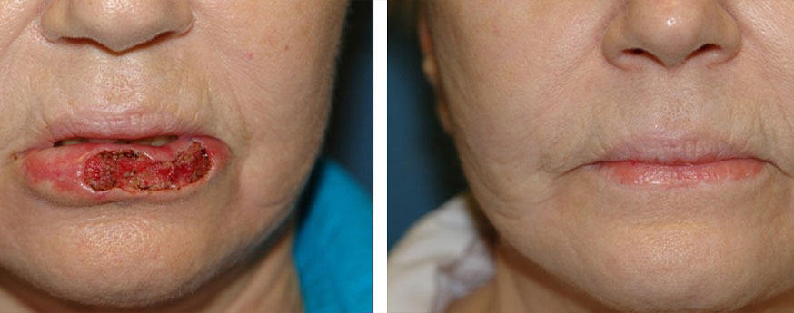
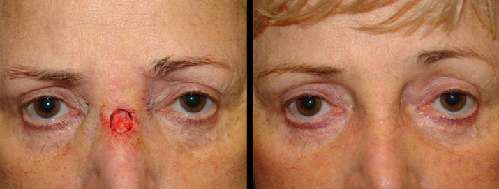
Skin Cancer
Before & Afters
*Each patient is unique, and individual results may vary.
See More Before & Afters
The Three Major Types of Skin Cancer
Basal Cell Carcinoma
Basal cell carcinoma is primarily found on the face or other exposed areas of the body. It is usually raised, translucent, and pink, with pearly borders, and may crust or bleed as it enlarges. It has a tendency to grow very slowly and invade local structures such as the nose, lips, or eyes. It almost never spreads (metastasizes) to any distant areas of the body but can cause significant local damage if not treated early. Early surgical cure is almost 99% effective.
Squamous Cell Carcinoma
Squamous cell carcinoma is usually found on exposed areas of the body, such as the scalp, ears, and lips, but can occur elsewhere. It is usually raised, pink, and has opaque patches that ulcerate or become crusty in the center. It has a greater tendency to metastasize than basal cell carcinoma, but again, if treated early, it has an excellent chance of a complete cure.
Melanoma
Melanoma may arise in any area of the body. It is usually a brown-black or multicolored patch or plaque with an irregular border. It may originate in a preexisting mole or as an isolated lesion. Any change in the appearance of a mole is highly suspicious. Melanoma has a high rate of metastasis if not treated early and is perhaps the most dangerous skin cancer.
If you have any moles or skin growths that you are concerned about, it is best to have your doctor examine these. Any growth that is suspicious should be biopsied to rule out the possibility of cancer. Remember, if treated early, almost all skin cancers are curable.
The Removal and Destruction of Skin Cancer
MOHS Micrographic Surgery
MOHS surgery is a modified form of surgical excision that provides an accurate assessment of the completeness of tumor removal and, as a result, has a very high cure rate and may be more tissue-sparing than conventional surgery. MOHS surgery is usually reserved for those instances where it is very important to preserve normal skin (i.e., eyes, nose, lips, ears, etc.) or where other types of treatments have either failed or would not be as successful. Call to learn more about our treatment methods for skin cancer in Gulf Shores today.

Electrodesiccation and Curettage (ED&C)
This form of destruction of cancer essentially consists of scraping (curettage) and burning with electrical current (electrodesiccation) the visible and palpable tumor and some surrounding skin. This procedure does not provide a method of assessing whether the tumor is completely destroyed. It usually results in a circular wound that heals with a circular scar in 3-8 weeks. It should be used only to treat primary (never treated) skin cancer and not on certain body sites.
Surgical Excision
This method provides for the removal of skin cancer and subsequent repair of the wound thus created. It provides tissue for microscopic assessment of the completeness of tumor removal. Surgical excision usually heals in 1-2 weeks with a linear or geometric scar, depending on the extent of surgery required. However, some patients, depending on the nature of their tumor, could require extensive reconstruction. After the scars are mature (usually 12- 18 months), additional plastic surgical techniques may be used to improve and/or camouflage them.
Cryosurgery
A form of destruction of skin cancer utilizing intense cold in the form of liquid nitrogen. Like ED&C, this method does not provide for assessment of complete tumor destruction. Ideally, it should be performed with the use of cryoprobes (needles in the skin used to measure temperature changes) in order to obtain optimal destruction of the tumor. It usually heals, similar to electrodesiccation and curettage.
Radiation Therapy
A form of destruction of skin cancer utilizing specifically controlled radiation energy. It is useful for those patients who would not tolerate surgical procedures either because of medical problems or because of fear of surgery. It is also useful in those anatomic locations that would necessitate extensive reconstruction with other modalities. It can be used to treat primary tumors. However, it does not provide an assessment of the completeness of tumor destruction.

MOHS Micrographic Surgery
Experience confirms that the most accurate method of removing skin cancers and preserving the most adjacent tissue is a technique known as “MOHS Micrographic Surgery.”
Approximately 40 years ago, Dr. Frederic MOHS at the University of Wisconsin developed this technique for the removal of skin cancers. This technique offers patients the highest chance of cure with maximal preservation of normal tissue, thereby reducing the difficulty of reconstruction of defects that result from tumor removal. However, because this method is time-consuming, requires highly specialized training, and is not always necessary for treating skin cancer, few dermatologists in the United States are equipped to offer such treatment.
Steps Involved in MOHS Micrographic Surgery:
- Surgical removal (debulking) of the visible portion of the skin cancer with curettage (scraping) or excision.
- The surgical removal of a thin layer of tissue at the bed and periphery of the cancer with meticulous mapping and color coding of the tissue
- The examination of this excised tissue immediately under the microscope, using the mapping to determine the extent of the tumor and the need for surgery further.
- If residual cancer is detected, we are able to locate remaining cancer and steps 2 through 3 are repeated until the tumor is completely removed.
MOHS Micrographic Surgery: What to Expect
This surgery is usually performed under local anesthesia as an outpatient. The actual surgery takes 15-30 minutes per stage of tissue removal, after which a temporary bandage is placed on the wound. The excised tissue is then prepared for microscopic evaluation in a process that may take up to 11/2 hours. During this time, you may wait in the waiting room.
If the examination of the tissue removed reveals that your tissue still contains cancer cells, the procedure will be repeated as soon as possible. Several excisions and microscopic exams may need to be done in one day and rarely may require two days. The average number of surgical stages for most skin cancers is two or three, so most patients have their entire skin cancer removed by midday on the day of surgery. Plan on spending the entire day and bring something to do or read.

The Surgical Wound
When the skin cancer is completely removed, a decision is then made with regard to the appropriate method for treating the wound that has been created. The usual choices include:
- Letting the wound heal by itself (granulation)
- Closing the wound with stitches (primary repair)
- Closing the wound with a flap (moving skin around)
- Closing the wound with a graft (transplanting skin from one body site to another)
- Delayed closure of the wound with the above choices.
The method used will be determined by the nature, extent, and location of the tumor and the resultant wound. This most appropriate method of repair is usually apparent before performing MOHS surgery, but on occasion, the exact nature and extent of the tumor and the resultant wound are not apparent until after surgery. Thus, occasionally, the decision as to how to repair the wound may not be possible until after the surgery is performed, and sometimes, the initial treatment plan may have to be altered.

Schedule Your Consultation
With Us Today
At McCollough Plastic Surgery, we bring a wealth of experience and expertise to skin cancer treatments, such as MOHS Micrographic Surgery. Contact us today to meet with a renowned expert in helping patients reclaim their aesthetics and general quality of life after cancer. For more information, schedule your consultation to discuss skin cancer in Gulf Shores with our plastic surgeons.